What are epidermal cysts / follicular infundibular cysts / epidermal inclusion cysts / epidermoid cysts ?
Epidermal cysts are also called follicular infundibular cysts, epidermoid cysts, and epidermal inclusion cysts. They are formed as a result of the implantation of epidermal elements in the dermis. Their centers are filled with keratin and lipid-rich fragments of epidermal cells. These cysts are benign skin cysts, with centers containing soft, cheese-like material. They are always attached to the skin.
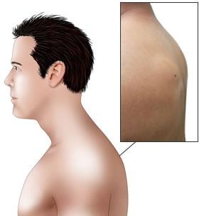
Epidermal cysts usually grow on the face, ear, neck, and back. They range from a few millimeters to a few centimeters. There is often a punctum (black dot) on their surface. When they become bigger, they may raise, rupture with a pasty white discharge, or become infected with pus collection or pus discharge.
Cause
Epidermal cysts are usually associated with blocked excretory ducts of the hair follicle or after skin piercing. The formation of epidermal cysts may be due to the following reasons:
- During embryonic life, epidermal cells remain below the surface of the skin and gradually grow and increase in size.
- Occlusion of the pilosebaceous unit
- Skin impact, skin puncture, or skin trauma during surgery, or the implantation of skin cells into deeper layers
- Human papillomavirus (HPV) infection
- Ultraviolet irradiation
- Occlusion of eccrine ducts
- Certain genetic syndromes are associated with epidermoid cysts. Such syndromes include Gardner's disease, basal cell nevus syndrome, and pachyonychia congenita
Symptoms
Treatment
Epidermal cysts should be treated with surgical excision. If there is infection, abscess formation, or pus discharge, the treatment choice is incision and drainage of pus, excision of the abscess wall, daily dressing, and possibly delayed closure of the wound. These surgeries can be done under local anesthesia in the clinic or under general anesthesia in a hospital. Please consult your surgeon for advice.
Traditional Epidermal Cyst Excision
An eye-shaped incision is made around the epidermal cyst. After the epidermal cyst is excised, the wound is closed with sutures. Please consult your surgeon for advice.
New Minimal Incision Epidermal Cyst Excision
A stab incision of 2-3 mm is placed over the epidermal cyst. The cyst content and cyst are completely removed via the small 2-3 mm wound. The wound is closed with micro-sutures. This will create the smallest wound and best cosmesis. Please consult your surgeon for advice.
FAQs
| Skin Polyps | Epidermal Cyst | |
|---|---|---|
| Shape | Irregular pedunculated skin nodules | Roundish nodule below skin surface. A punctum (small black dot) may appear on the surface. |
| Discharge | No discharge upon compression | May have some white discharge upon pressure |


